Microscopes
Royal Rife designed five models of microscopes, as well as equipment for taking photomicrographs. This page presents images of these instruments in approximate date order. Reproductions of the few extant photomicrographs of organisms follow the microscope equipment, along with a modern comparative photo and video.
Click on any image for larger version, which will open in a new page.
Early photomicrographic instrument & #1 microscope, 1920s - 1931
Rife with early photomicrographic instrument, date unknown.
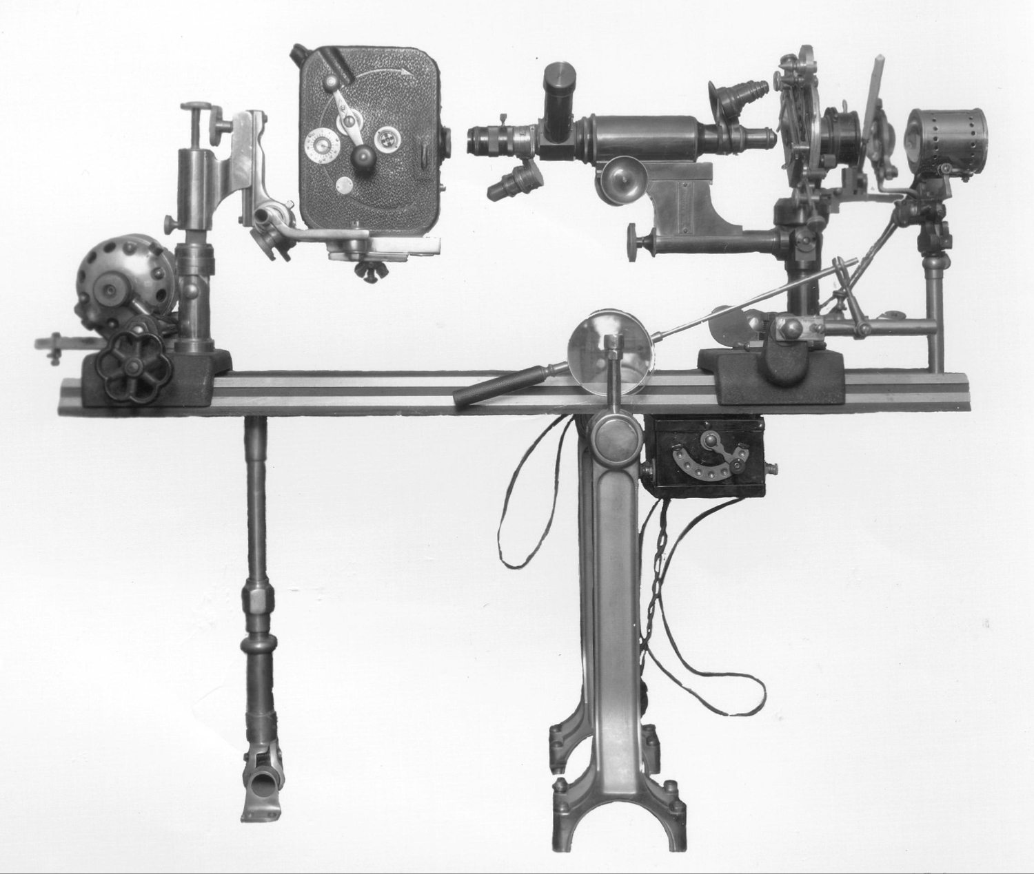
#1 Microscope. Horizontal microscope to the right, photography equipment to the left.
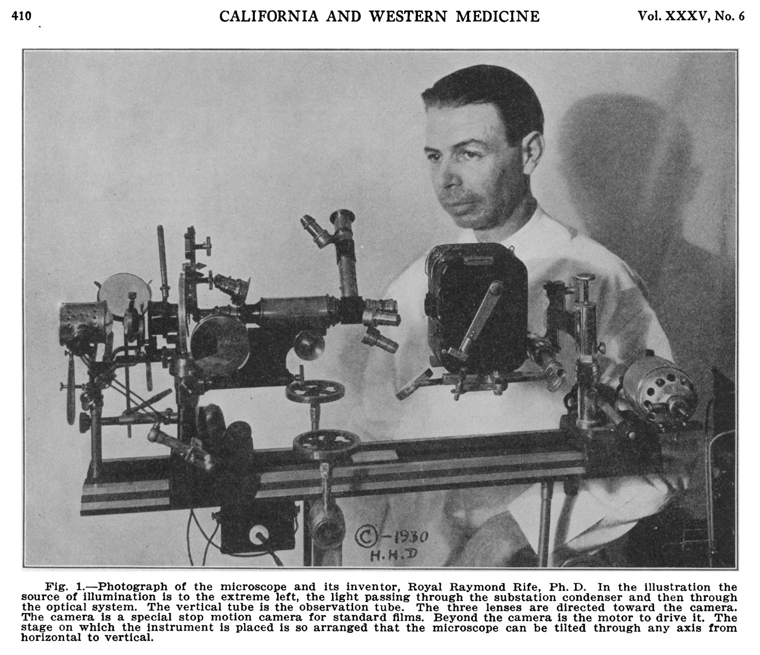
#1 Microscope, from California & Western Medicine journal article, Dec. 1931. Photography equipment to the right.
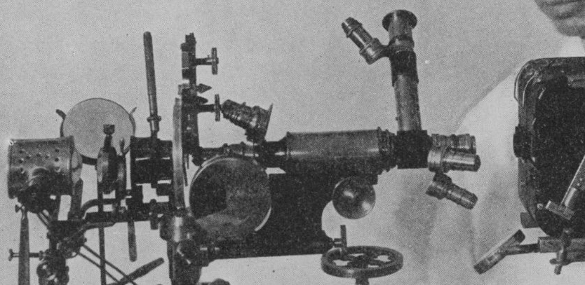
Previous photo from California & Western article, enlarged. Shows Rife's use of objectives as oculars.
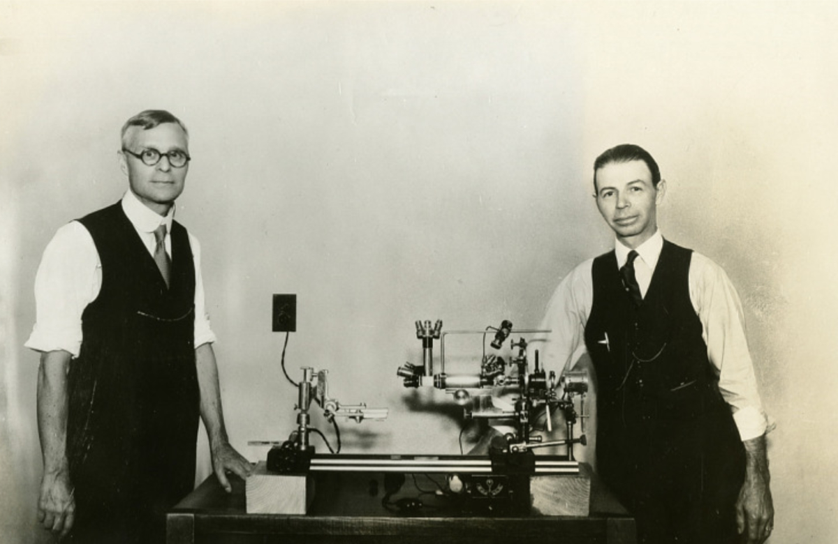
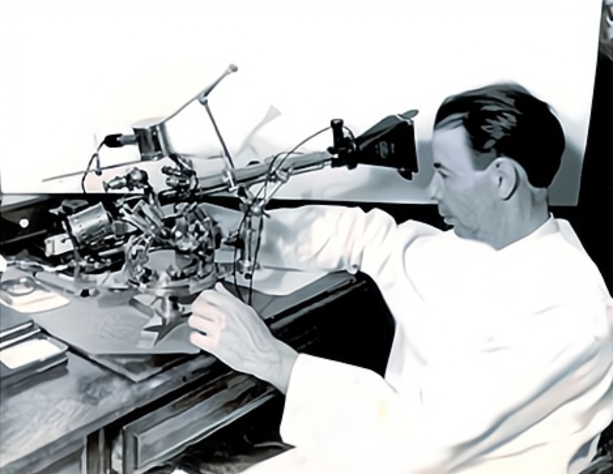
Rife & Kendall with #1 Microscope, from Smithsonian collection. Photography equipment removed.
Horizontal microscope, from Graphic Magazine article 30 January 1932. Possible transitional model.
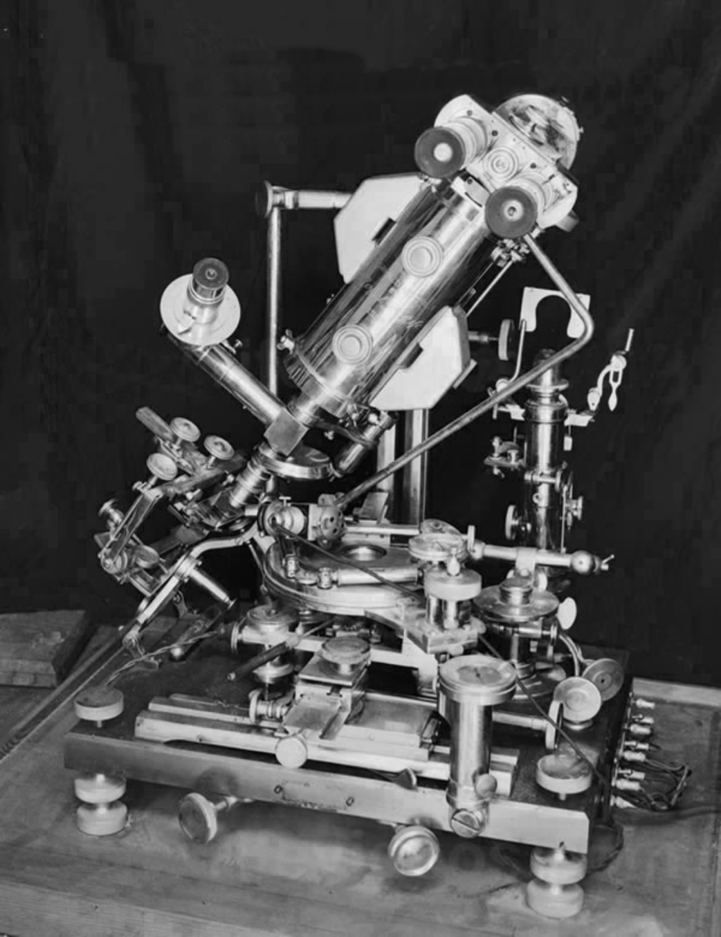
Microscope #2, 1932

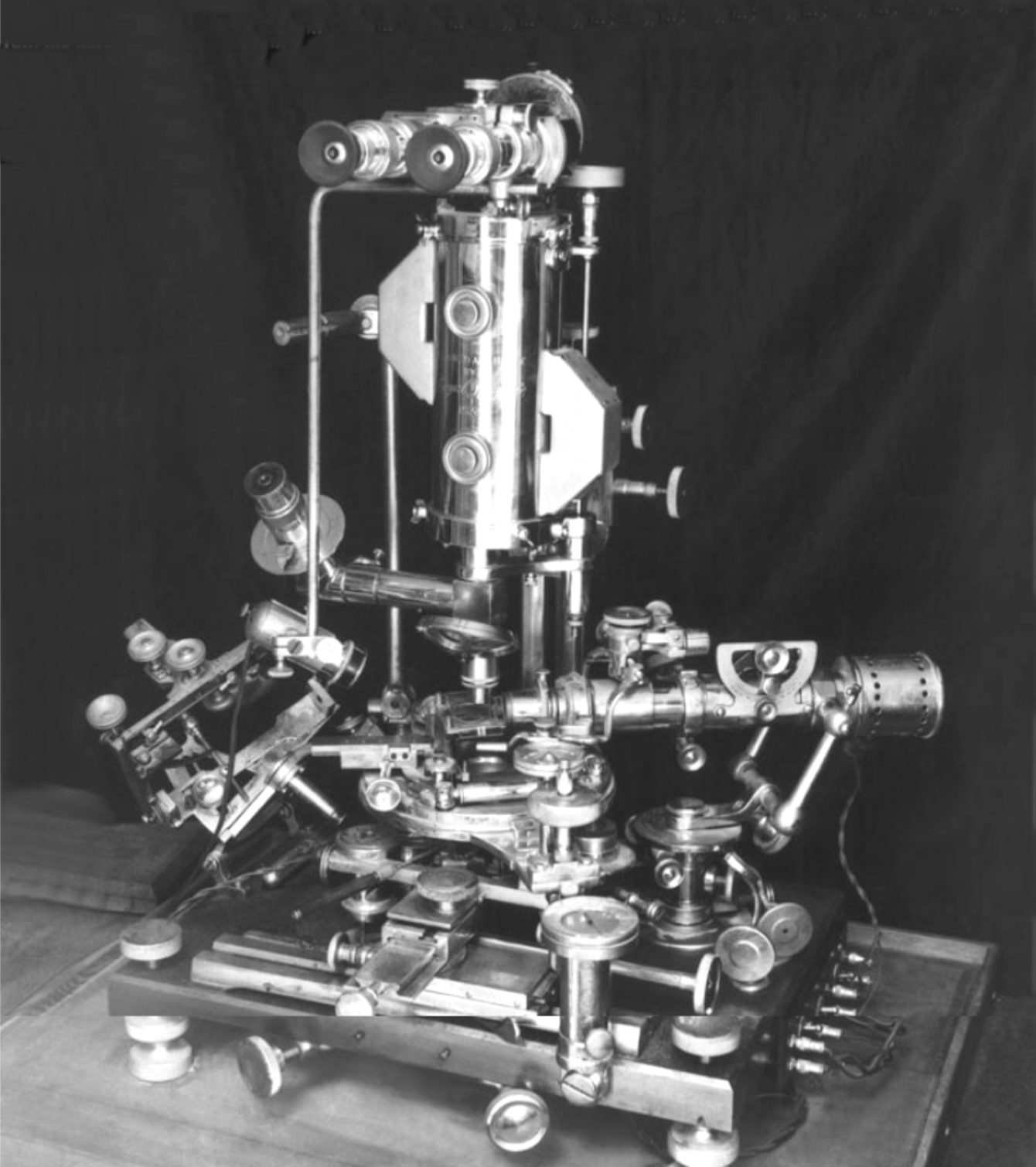
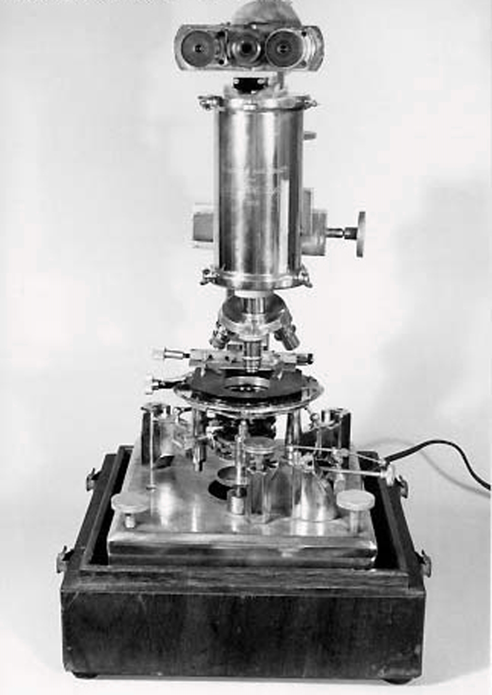
#2 Microscope, 1932.
#2 type of microscope, with illumination lamp above table.
#2 Rife 1932 microscope from Bonham auction, with illumination lamp below table. Parts missing.
#2 Rife 1932 microscope barrel from Bonham auction.
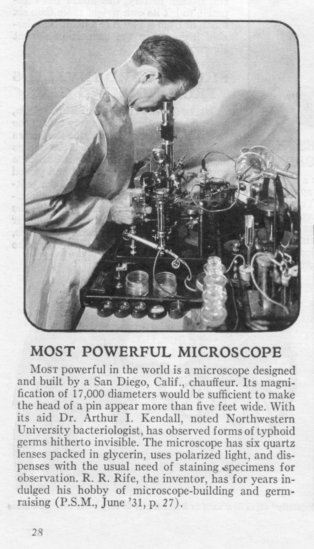
Rife with #2 Microscope in lab setting, from Popular Science February 1932.
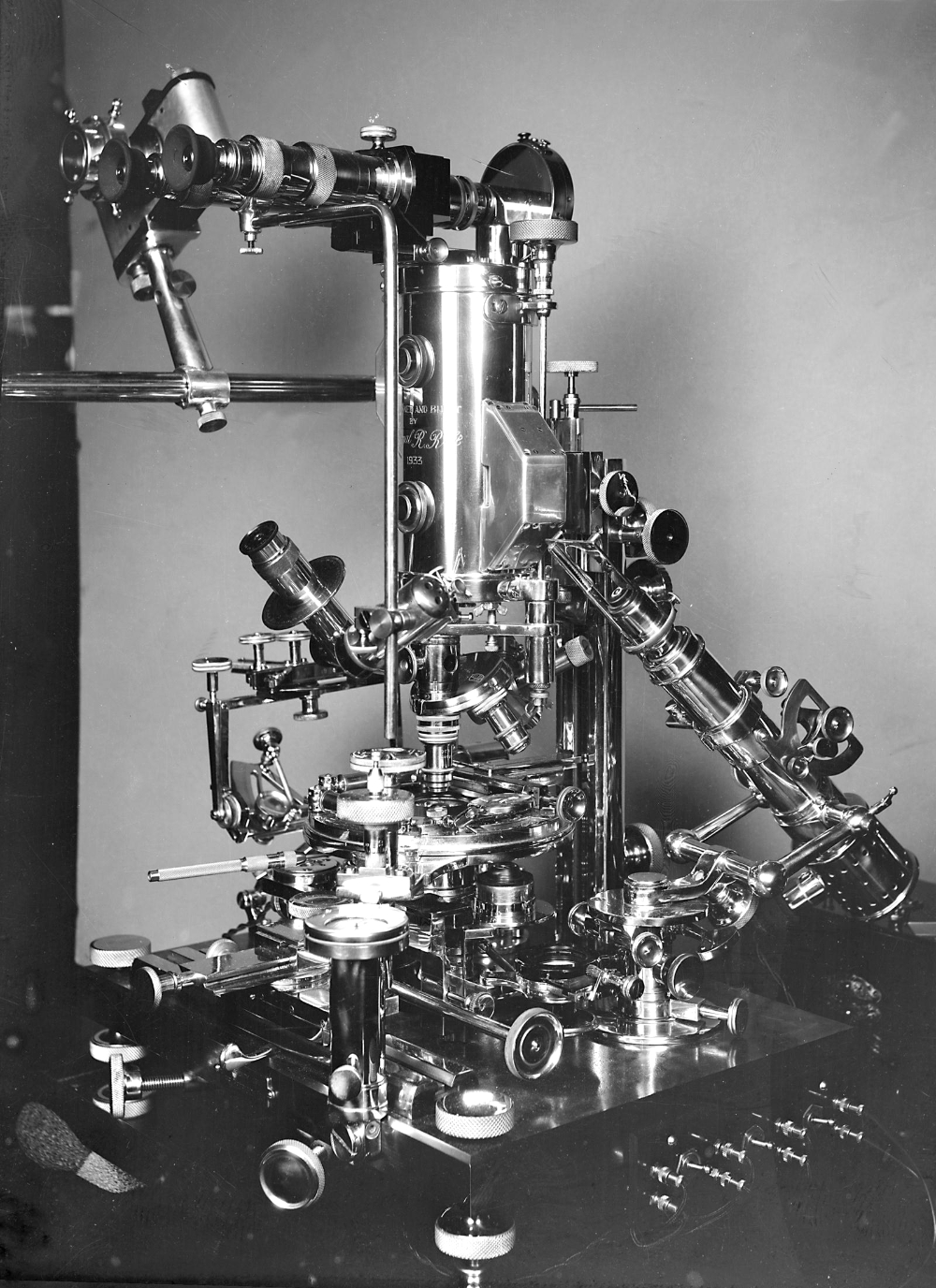
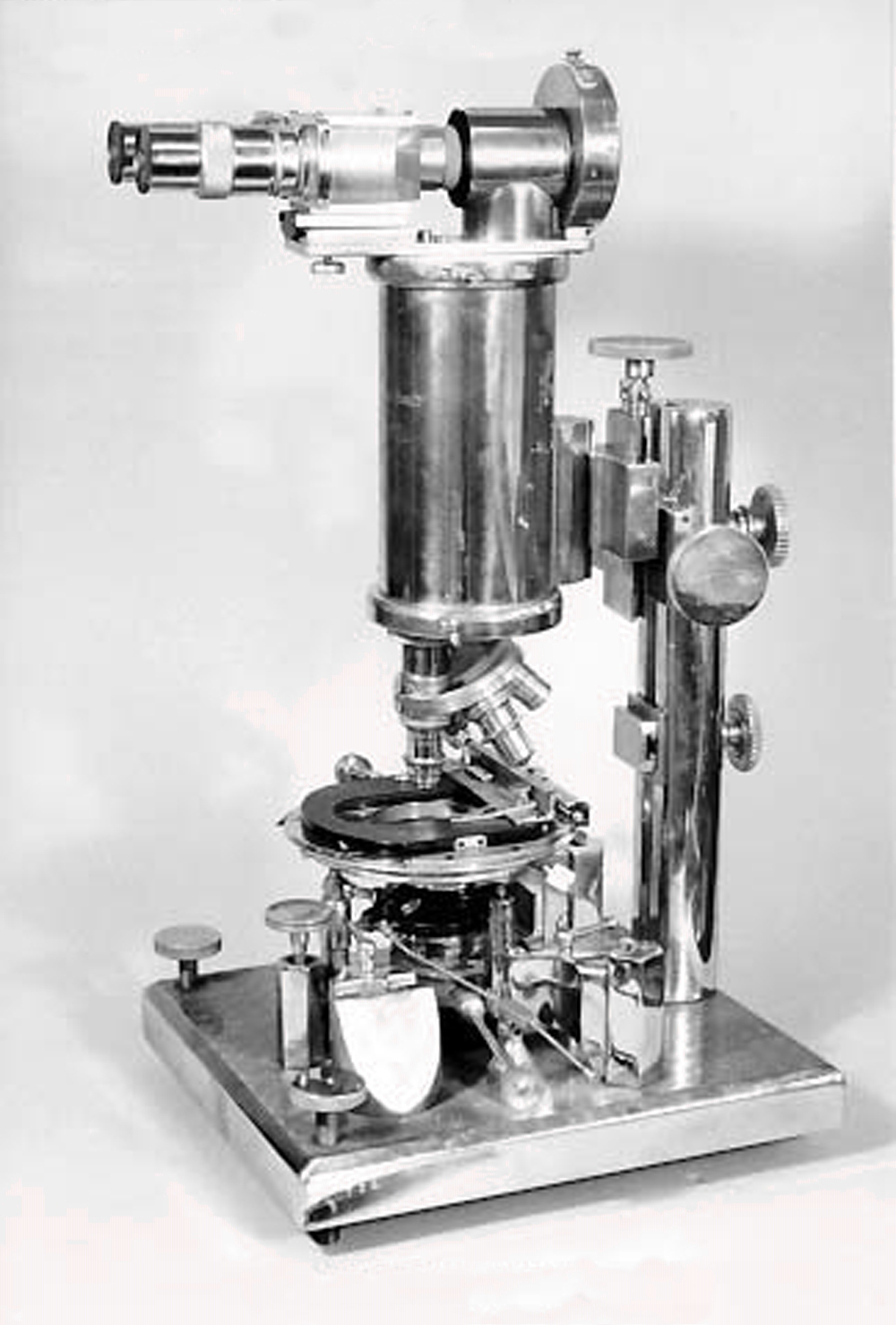
Microscope #3, 1933 (“Universal”)
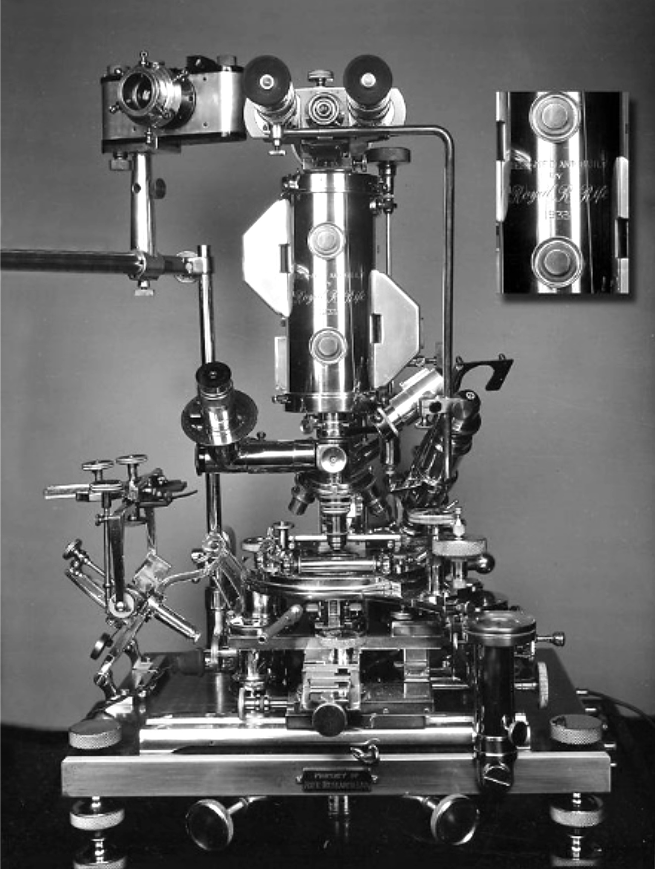
#3 Microscope (1933), with slit ultra attachment (right) in proper horizontal position for viewing.
#3 Microscope (1933), frontal view.
#3 Microscope (1933), side view 1.
#3 Microscope (1933), side view 2.
#3 Microscope (1933), with barrel and some adjusters tilted right.
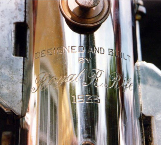
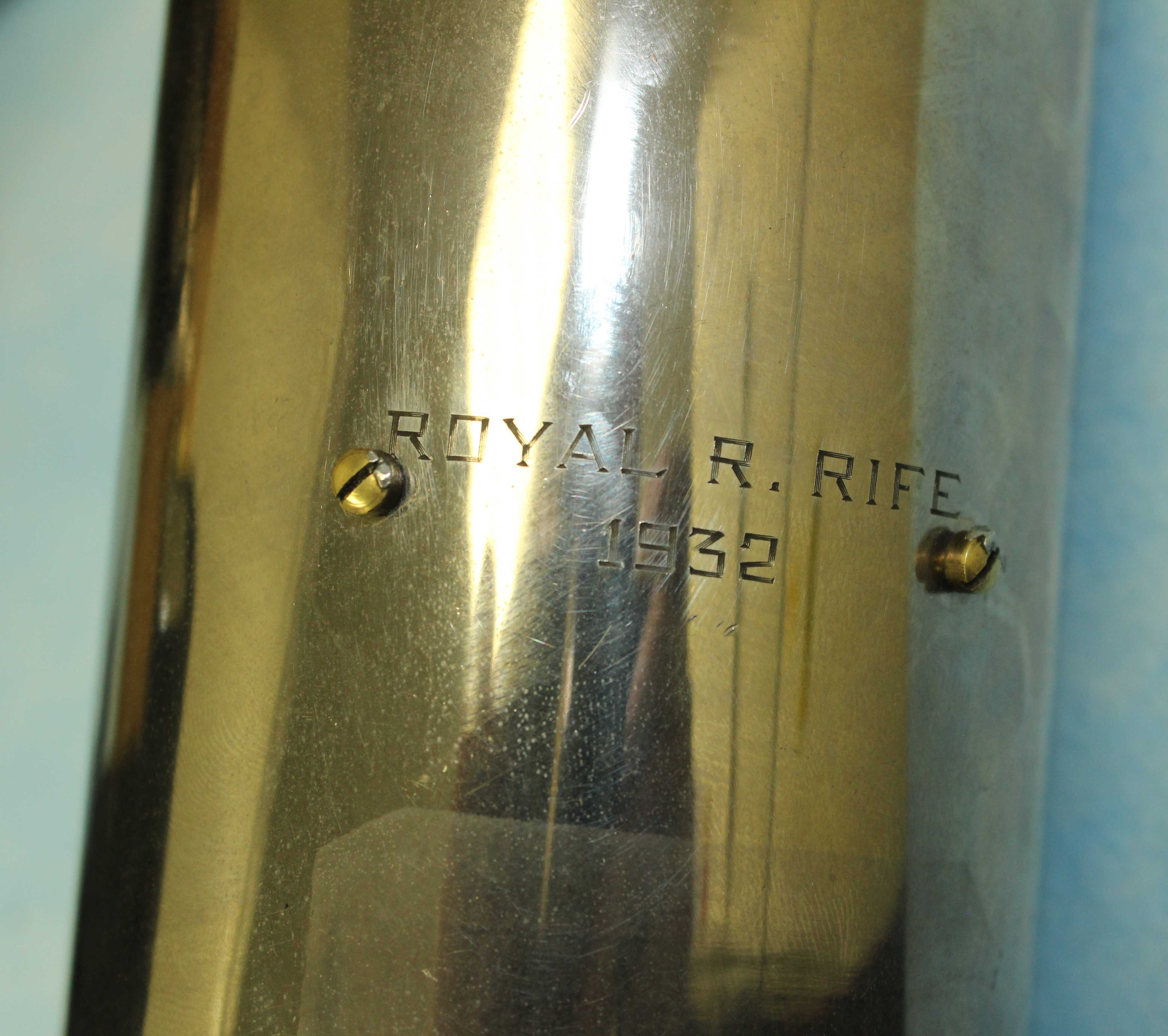
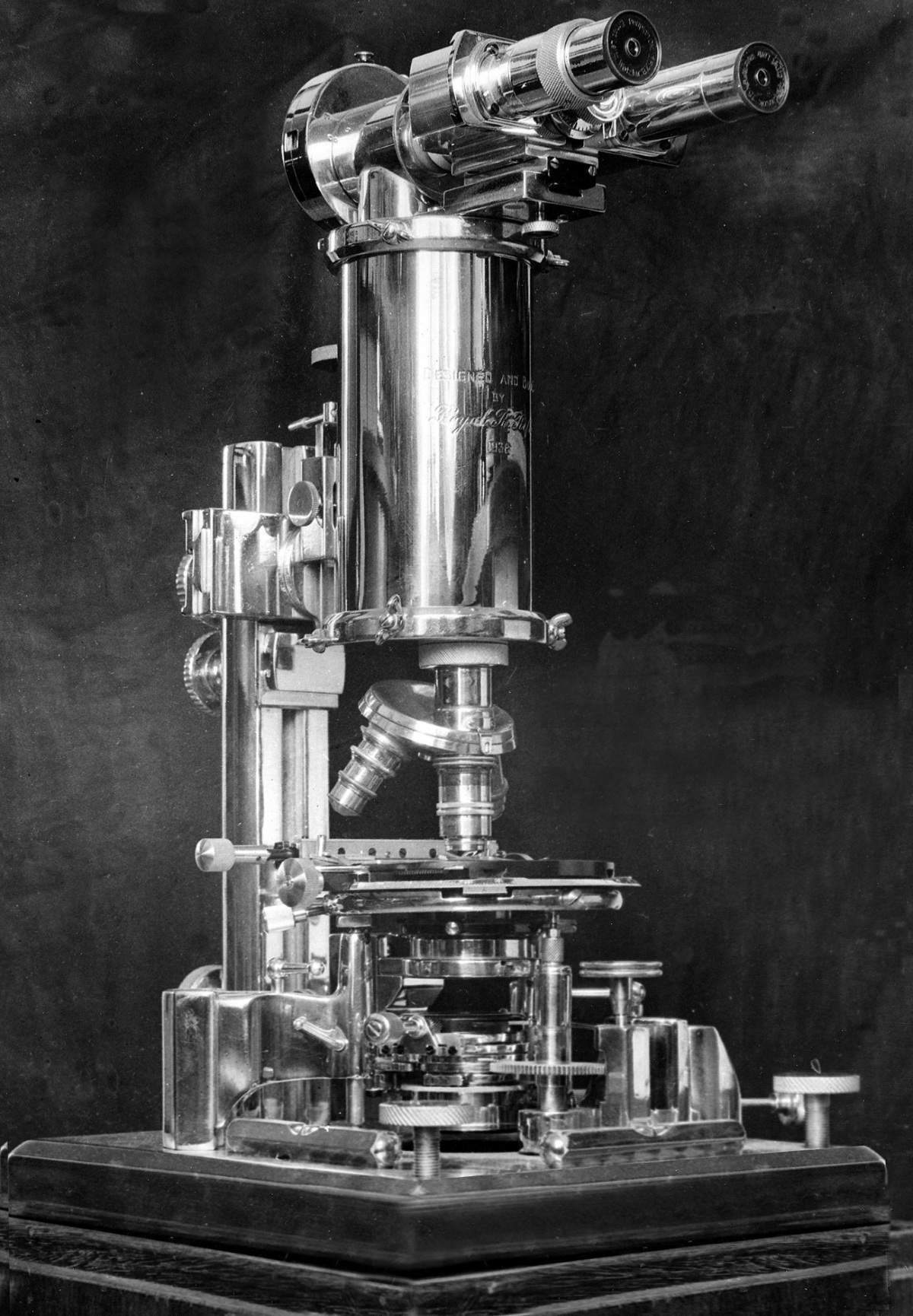
#3 Microscope (1933), barrel with inscription.
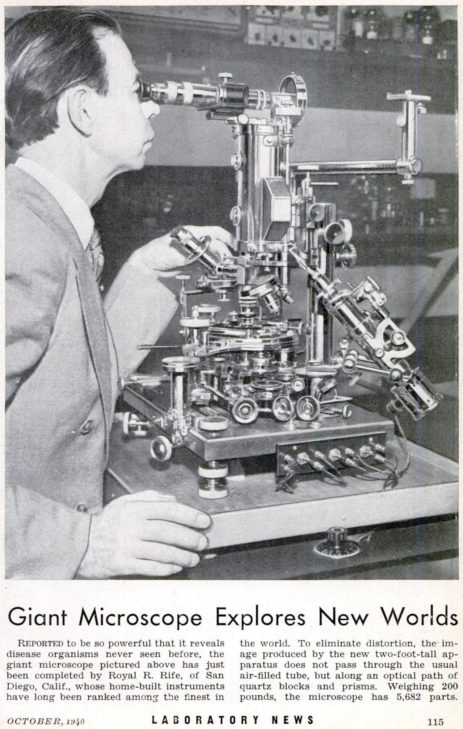
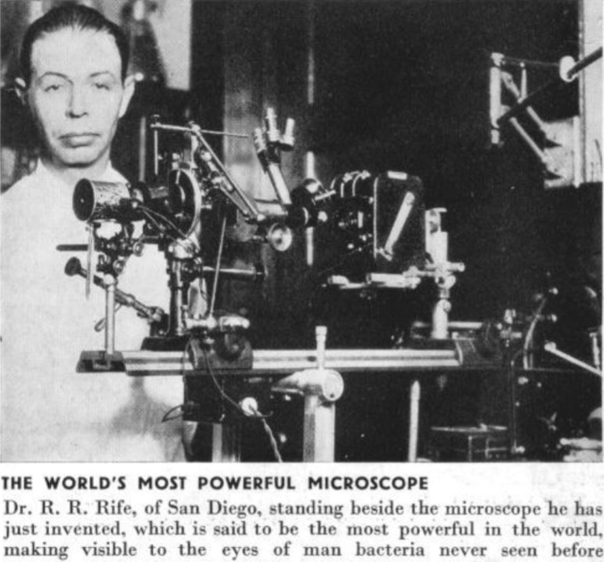
Rife with #3 Microscope, from Popular Science October 1940.
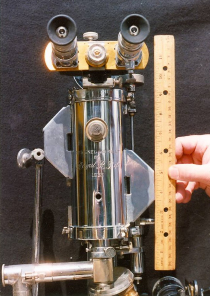
#3 Microscope (1933) partial, showing dimensions.
Microscope #4, 1935
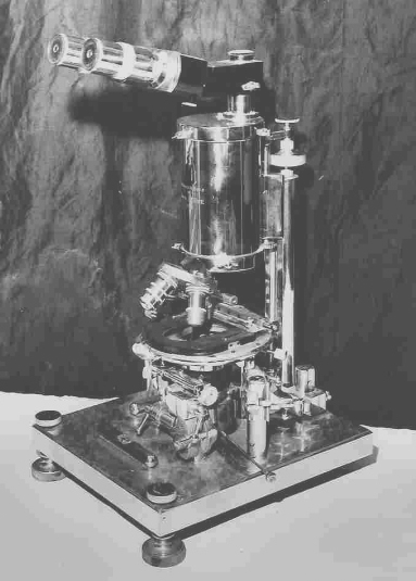
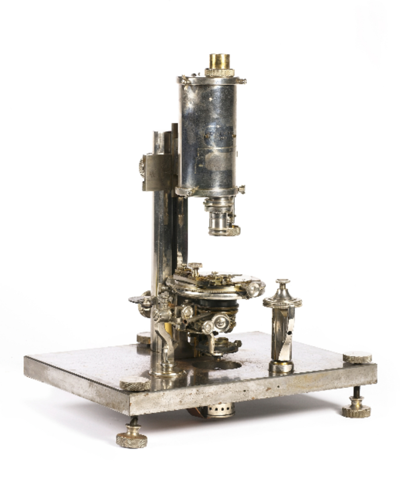
Purported #4 Microscope (1935).
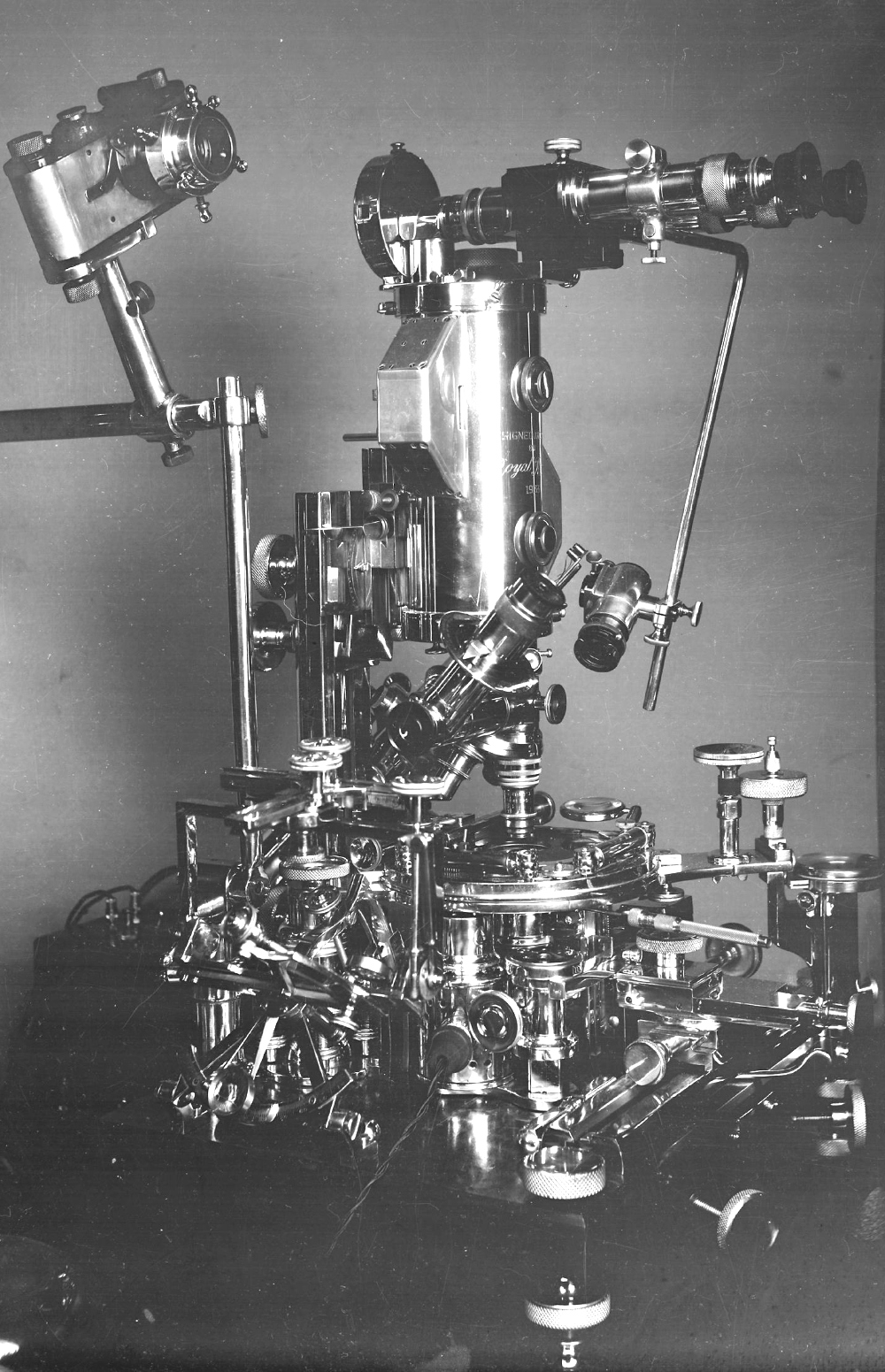
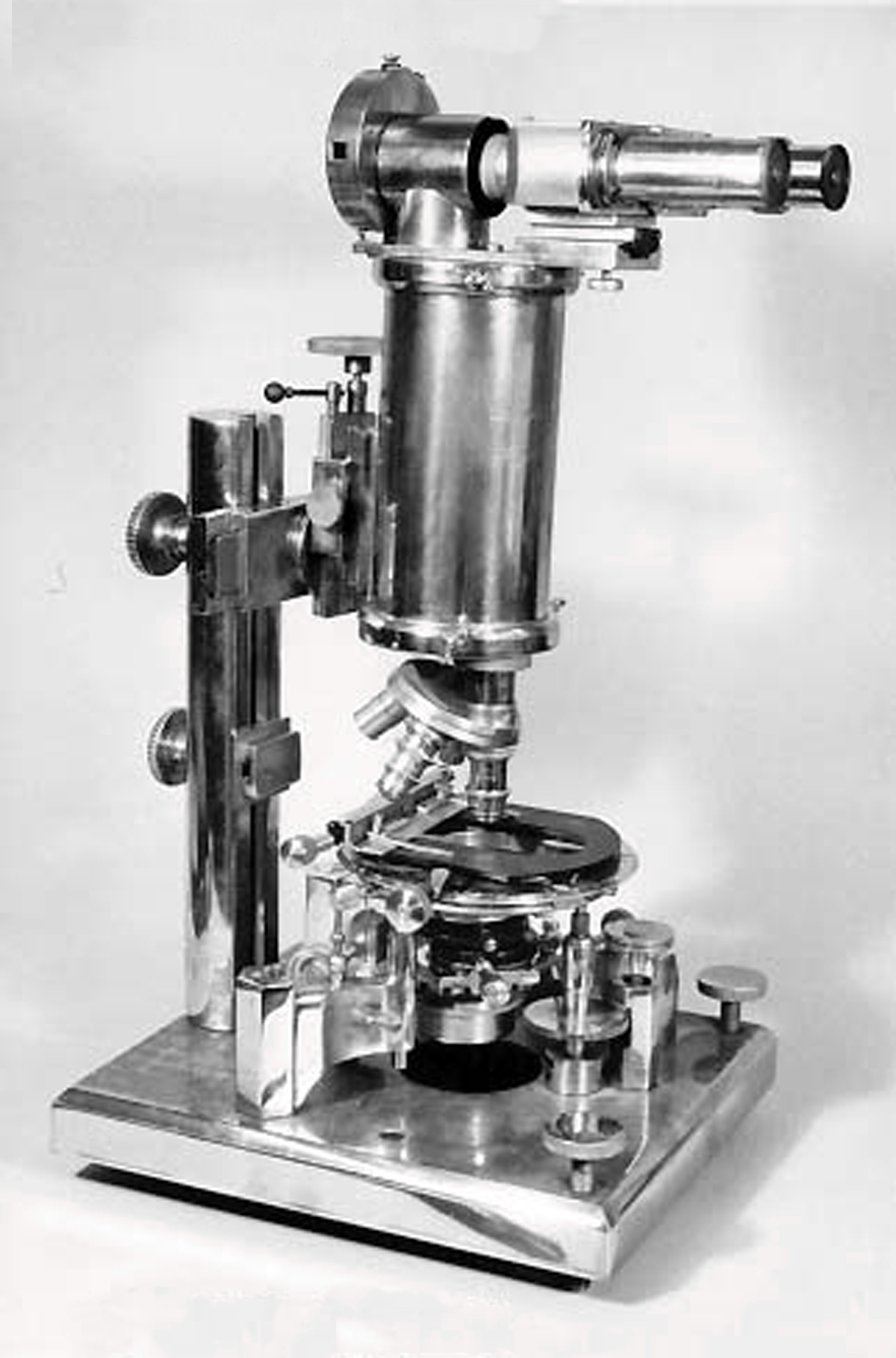
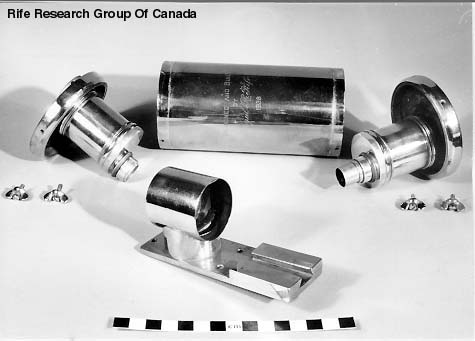
Microscope #5, 1938
#5 Microscope (1938).
#5 Microscope (1938), front view.
#5 Microscope (1938), side view 1.
#5 Microscope (1938), side view 2.
#5 Microscope (1938), substage optical components.
#5 Microscope (1938), barrel and optical components.
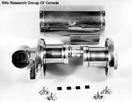
#5 Microscope (1938), barrel and optical components assembled.
Later photomicroscopic instrument
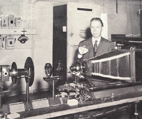
Rife with photomicroscope, probably early 1940s.
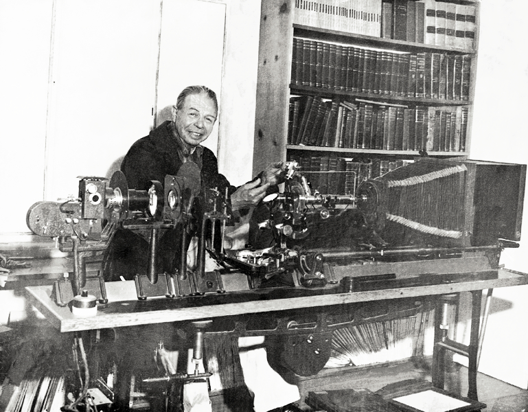
Rife with photomicroscope, circa 1953.
Rife’s patented lamp & the Risley prisms
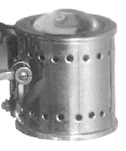
Closeup of lamp from #1 horizontal microscope image, rotated to vertical with convex lens facing up.
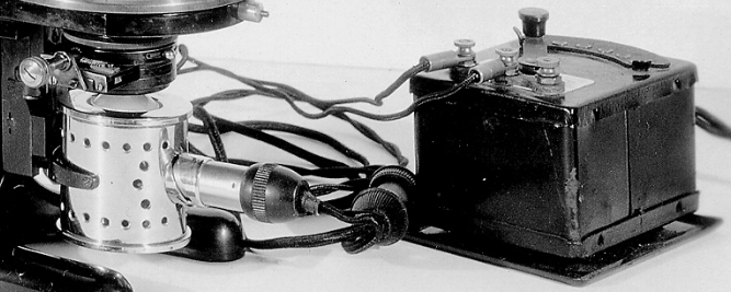
Closeup of Rife microscope lamp and power (rheostat) adjustment box installed on Otto Himmler microscope (see full image below).
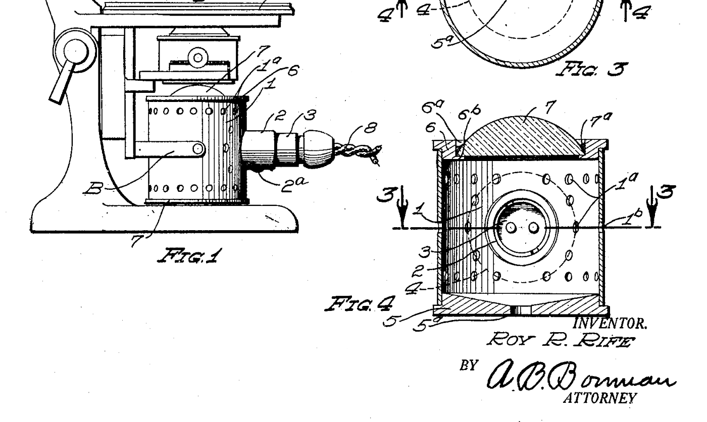
Closeup of Rife lamp drawing from his 1929 patent, #1727618. See full document at "Technical and Patents" section of website.
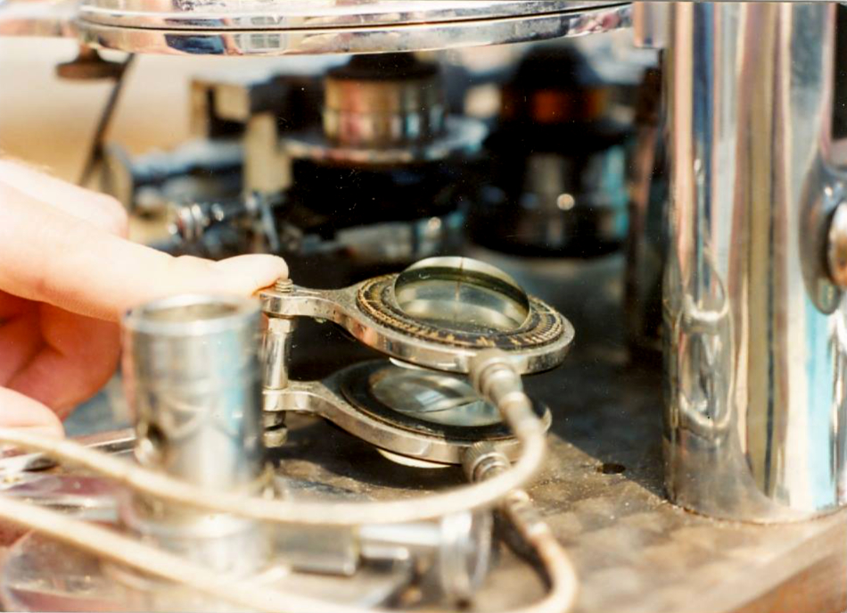
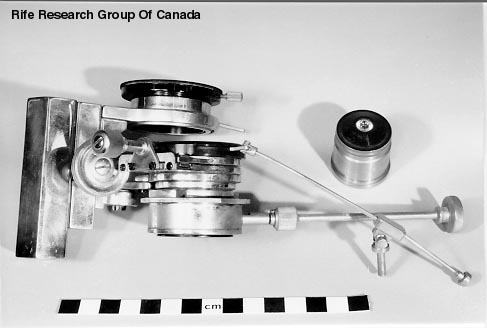
Two Risley prisms, from the #3 Rife microscope (1933). Each prism has two wedges.
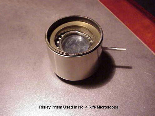
Risley single prism in its holder, from #4 Rife microscope (1935).

Risley single prism from #4 Rife microscope (1935), top wedge visible.
Other microscopes
Rife with a special design Leitz petrographic microscope. Used for finding virus colors and their mortal oscillatory rates (MORs), and other uses.
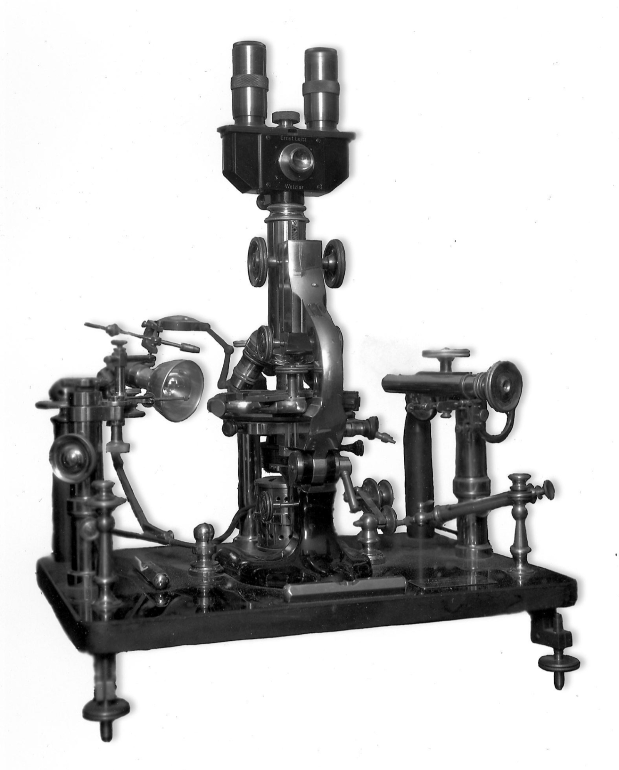
Microscope By Otto Himmler, Berlin, 190x. Retrofitted with Rife lamp. Owner unknown.
Photomicrographs
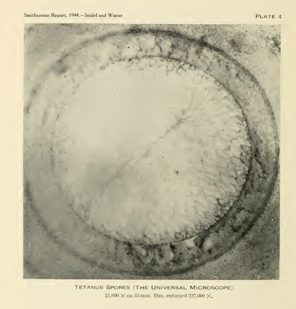
Tetanus spore photomicrograph, from Smithsonian report 1944.
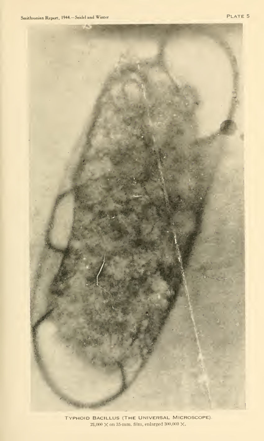
Typhoid bacillus photomicrograph, from Smithsonian report 1944.
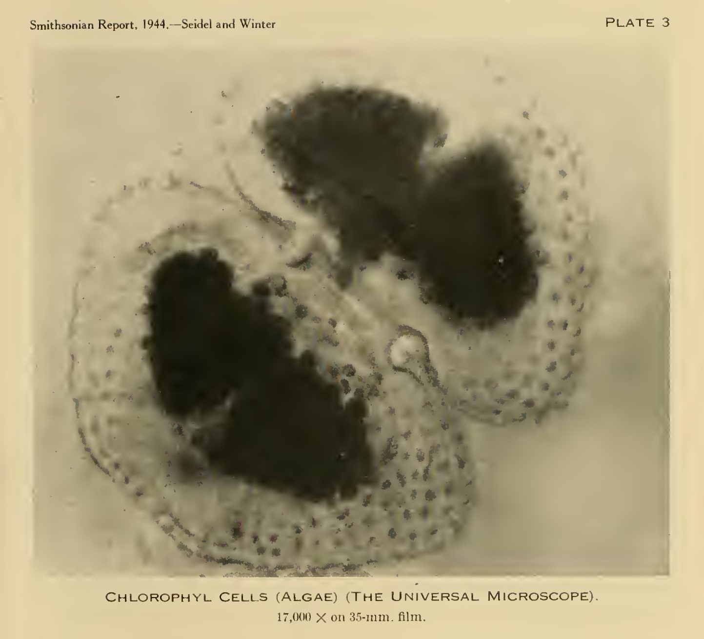
Chlorophyll cell photomicrograph, from Smithsonian report 1944.
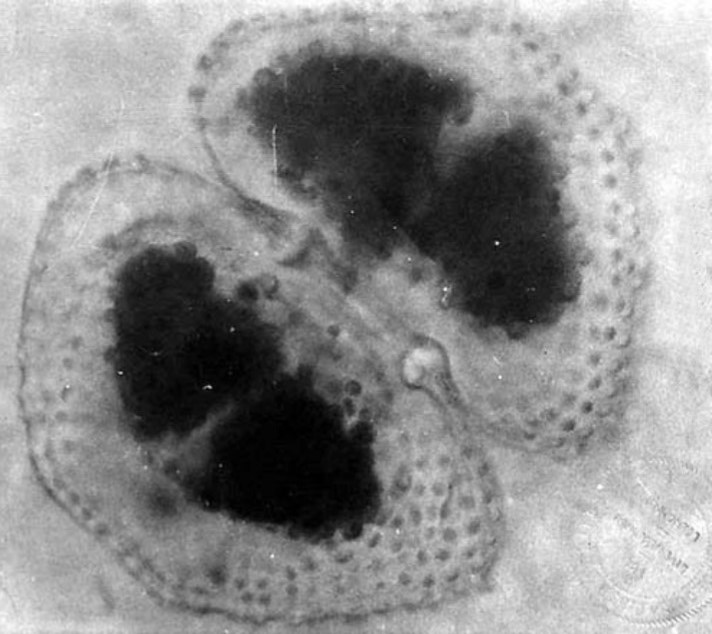
Chlorophyll cell photomicrograph 2, black & white with faint lab stamp in lower left corner.
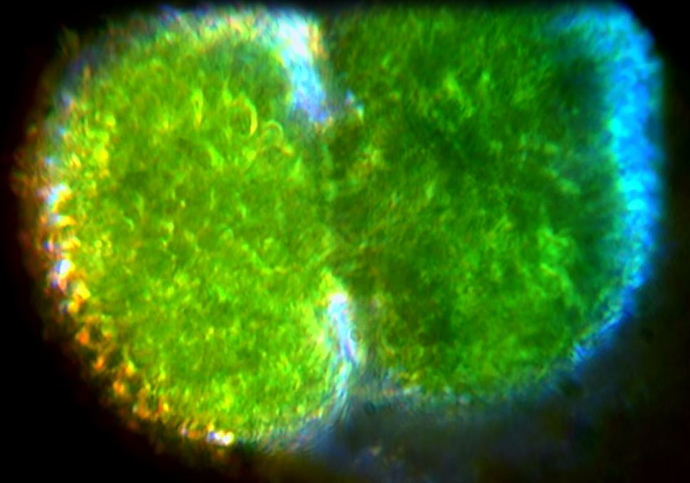
Modern comparison of live chlorophyll cell photo, from Truman microscope video (see below). 100x oil immersion objective, with Risley prism.
Chlorophyll live cell viewed with Truman microscope, 100x oil immersion objective with Risley prism.